The human body
The human eye
The core of each eye lens forms around birth and is never replaced, and the retina holds about 120 million rods and 6 million cones.

At a glance
120 million rods
Rod photoreceptors in the human retina, about
6 million cones
Cone photoreceptors in the human retina, about
1.5 years
Average uncertainty when a lens core dates a birth year
200 degrees
Width of the field of view, about
120 days
Typical life of a red blood cell
A lens core as old as you are
Most of the protein in the core (nucleus) of the human eye lens forms around birth. It is never subject to turnover or remodelling once formed, so the core is about as old as the person.
Scientists can date it with carbon-14. Above-ground nuclear tests between 1955 and 1963 left a spike of carbon-14 in the atmosphere, and the lens core keeps the carbon it took in at the time it formed.
In a test on 13 lenses from 13 deceased people, a lens core gave a person's birth year to within an average of 1.5 years at 95% confidence, excluding one case.
In another set of 14 donors born between 1948 and 1993, the carbon-14 in the lipids of the lens core matched the level in the air in each donor's birth year.
The lens does not lose and replace its cells over a lifetime. It grows by adding fibre cells.
A lipid called sphingomyelin also persists in the lens core. Lipids normally turn over quickly, and this was reported as the first evidence of a lipid that lives longer than the others in the body.
How light is brought into focus
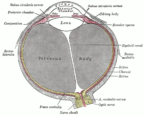
Light enters through the pupil, and the iris controls how much of it the pupil lets in.
The cornea is the clear, dome-shaped front layer. It bends light, and the lens works with it to focus light onto the retina.
For distance vision the ciliary muscle is relaxed, the zonule fibres are taut and the lens is held flatter.
To focus on something near, the ciliary muscle contracts and the zonule fibres relax. The lens becomes more spherical and its refractive power rises.
The retina is the light-sensitive layer at the back of the eye. Its photoreceptors turn light into electrical signals that travel along the optic nerve to the brain.
The human field of view is about 200 degrees wide and 135 degrees tall.
Rods, cones and the sharpest spot
The human retina holds about 120 million rods and about 6 million cones.
Rods make up about 95% of the photoreceptors. They are the main drivers of vision in low light but cannot detect colour.
Most people have three kinds of cone, which respond to short (blue), medium (green) and long (red) wavelengths. The brain compares their signals to produce colour.
The fovea, the central region of the retina that gives the clearest vision, contains only cones, packed more closely than anywhere else.
Most cones sit in one place on the retina, the macula.
Where the optic nerve leaves the eye there are no photoreceptors, so an image that falls there is not seen. This is the blind spot.
Young and old parts of one body
Red blood cells typically live for about 120 days.
Fat cells are replaced at a rate of about 8% a year, so half of them are replaced in about 8 years.
The lens core does not follow this pattern of renewal. Its cells are not replaced, unlike red blood cells and fat cells.
Sources
- PLOS ONE, Radiocarbon dating of the human eye lens crystallines reveal proteins without carbon turnover throughout life
- ANSTO, Nuclear techniques confirm unique biology of human eye lens
- National Eye Institute, How the Eyes Work
- National Library of Medicine, NCBI Bookshelf, Anatomy, Head and Neck, Eye Ciliary Muscles
- Cleveland Clinic, Eyes: How They Work, Anatomy & Common Conditions
- University of Washington, Neuroscience For Kids: Retina
- Cleveland Clinic, Photoreceptors (Rods & Cones): Anatomy & Function
- University of Rochester Medical Center, What Are Red Blood Cells?
- Cell Biology by the Numbers, How quickly do different cells in the body replace themselves?